Labeled Areas Of The Back - Http Www Lamission Edu Lifesciences Alianat1 Chap1 Anatomical 20terminology Pdf : This image was chosen so you can see the anatomical areas without the organs.
Labeled Areas Of The Back - Http Www Lamission Edu Lifesciences Alianat1 Chap1 Anatomical 20terminology Pdf : This image was chosen so you can see the anatomical areas without the organs.. The muscles of the back are a group of strong, paired muscles that lie on the posterior aspect of the trunk they provide movements of the spine, stability to the trunk, as well as the coordination between the movements of the limbs and the back muscles are divided into two large groups: Like the cerebral cortex, it has two hemispheres. The shoulder is a complex combination of bones and joints where many muscles act to provide the widest range of motion of any part of the body. It contains the osteology, arthrology and myology of the spine and back. The lower back (where most back pain occurs) includes the five vertebrae in the lumbar region and supports much of the weight of the upper body.
It is the surface of the body opposite from the chest and the abdomen. Areas of the back : Numerous muscles help stabilize the three joints of. It provides several important functions, including: Protecting the spinal cord and nerves.
Deep Back Muscles Anatomy Innervation And Functions Kenhub from thumbor.kenhub.com About anatomy of the spine. Surface marking of kidney location. Hip anatomy, function and common problems front view of the hip joint bones. The back is the body region between the neck and the gluteal regions. The back functions are many, such as to house and protect the spinal cord, hold the body and head upright, and adjust the movements of the upper and lower limbs. Labeled areas of the back. The vertebral column houses the spinal canal, a cavity that. Nerves in your lower back five pairs of lumbar spinal nerves labeled l1 to l5 branch off your spinal cord and exit through small holes between the vertebrae.
The muscles of the back are a group of strong, paired muscles that lie on the posterior aspect of the trunk they provide movements of the spine, stability to the trunk, as well as the coordination between the movements of the limbs and the back muscles are divided into two large groups:
Hip anatomy, function and common problems front view of the hip joint bones. The anatomy of the hip and back is comprised of numerous parts that can be injured or wear out, and many problems that occur in this area can display the exact same symptoms or pathology. The spaces between the vertebrae are maintained by intervertebral discs that act like shock absorbers throughout the spinal column to cushion the bones as the body moves. The part of the nerve that emerges out of the spine is called the nerve root. On this page, you'll learn about each of these muscles, their locations and functional anatomy. The brain is one of the largest and most complex organs in the human body. Human anatomy · july 23, 2016. Labeled areas of the back. Ligaments hold the vertebrae in place, and tendons attach the muscles to the. Spinal anatomy and back pain. Together, the brain and spinal cord make up the central nervous system. The spine supports about half the weight of the body. Some of these muscles are quite large and cover broad areas.
The lower back (where most back pain occurs) includes the five vertebrae in the lumbar region and supports much of the weight of the upper body. It contains the osteology, arthrology and myology of the spine and back. It comprises the vertebral column (spine) and two compartments of back muscles; The vertebral column runs the length of the back and creates a central area of recession. The shoulder is a complex combination of bones and joints where many muscles act to provide the widest range of motion of any part of the body.
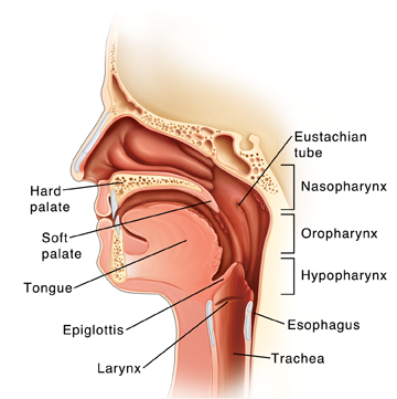
Parts Of The Throat And Neck Saint Luke S Health System from api.kramesstaywell.com About anatomy of the spine. Anatomy of the back spine and back muscles kenhub.the area directly behind a person: On this page, you'll learn about each of these muscles, their locations and functional anatomy. This human anatomy module is composed of diagrams, illustrations and 3d views of the back, cervical, thoracic and lumbar spinal areas as well as the various vertebrae. The muscles of the back are a group of strong, paired muscles that lie on the posterior aspect of the trunk they provide movements of the spine, stability to the trunk, as well as the coordination between the movements of the limbs and the back muscles are divided into two large groups: The lumbar and sacrum region make up the bone of the lower back anatomy. The vertebral column, also known as the backbone or spine, is part of the axial skeleton.the vertebral column is the defining characteristic of a vertebrate in which the notochord (a flexible rod of uniform composition) found in all chordates has been replaced by a segmented series of bone: Other muscles are small and cover much less space.
To put it plainly, sometimes hip pain comes from the hip, but a lot of times hip pain comes from the back.
The muscles of the back can be arranged into 3 categories based on their location: The vertebral column, also known as the backbone or spine, is part of the axial skeleton.the vertebral column is the defining characteristic of a vertebrate in which the notochord (a flexible rod of uniform composition) found in all chordates has been replaced by a segmented series of bone: The bones of the pelvis and lower back work together to support the body's weight, anchor the abdominal and hip muscles, and protect the delicate vital organs of the vertebral and abdominopelvic cavities. Bones of the pelvis and lower back. The back's muscles start at the top of the back (named the cervical vertebrae) and go to the tailbone (also named the coccyx). It provides several important functions, including: Like the cerebral cortex, it has two hemispheres. The extrinsic (superficial) back muscles, which lie most superficially on the back. Spinal anatomy is a remarkable combination of strong bones, flexible ligaments and tendons, large muscles and highly sensitive nerves. The spine's four sections, from top to bottom, are the cervical (neck), thoracic (abdomen,) lumbar (lower back), and sacral. Browse 384 human anatomy organs back view stock photos and images available, or start a new search to explore more stock photos and images. Spinal anatomy and back pain. Surface marking of kidney location.
Anatomy of the back spine and back muscles kenhub.the area directly behind a person: It comprises the vertebral column (spine) and two compartments of back muscles; It contains the osteology, arthrology and myology of the spine and back. It is the surface of the body opposite from the chest and the abdomen. Kidney location (anatomy) picture :
Pin On Anatomy And Physiology from i.pinimg.com It is the surface of the body opposite from the chest and the abdomen. Some of these muscles are quite large and cover broad areas. Like the cerebral cortex, it has two hemispheres. The back functions are many, such as to house and protect the spinal cord, hold the body and head upright, and adjust the movements of the upper and lower limbs. It is particularly interesting for physiotherapists, osteopaths, rheumatologists, neurosurgeons. Human anatomy · july 23, 2016. Spinal anatomy and back pain. This human anatomy module is composed of diagrams, illustrations and 3d views of the back, cervical, thoracic and lumbar spinal areas as well as the various vertebrae.
On this page, you'll learn about each of these muscles, their locations and functional anatomy.
Bones of the pelvis and lower back. The spaces between the vertebrae are maintained by intervertebral discs that act like shock absorbers throughout the spinal column to cushion the bones as the body moves. Superficial back muscles, intermediate back muscles and intrinsic back muscles.the intrinsic muscles are named as such because their embryological development begins in the back, oppose to the superficial and intermediate back muscles which develop elsewhere and are therefore classed as extrinsic muscles. Spinal anatomy is a remarkable combination of strong bones, flexible ligaments and tendons, large muscles and highly sensitive nerves. The spine's four sections, from top to bottom, are the cervical (neck), thoracic (abdomen,) lumbar (lower back), and sacral. Spinal anatomy and back pain. Like the cerebral cortex, it has two hemispheres. The latissimus dorsi originates from the lower part of the back, where it covers a wide area. The brain is one of the largest and most complex organs in the human body. Human anatomy · july 23, 2016. Browse 384 human anatomy organs back view stock photos and images available, or start a new search to explore more stock photos and images. It is made up of more than 100 billion nerves that communicate in trillions of connections called synapses. The anatomy of the hip and back is comprised of numerous parts that can be injured or wear out, and many problems that occur in this area can display the exact same symptoms or pathology.
This image was chosen so you can see the anatomical areas without the organs areas of the back. The vertebral column of the lower back includes the five lumbar vertebrae, the sacrum, and the coccyx.


0 Komentar